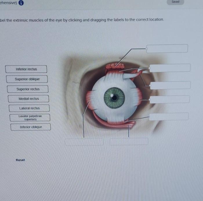
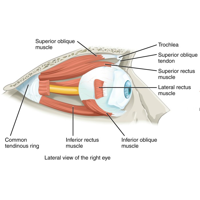
Label the extrinsic eye muscles. – Label the extrinsic eye muscles, a crucial aspect of ophthalmology, involves understanding the anatomy and function of the six muscles responsible for controlling eye movements. These muscles, innervated by cranial nerves, play a vital role in maintaining binocular vision, enabling precise eye coordination, and facilitating various eye movements essential for daily activities.
Delving into the intricate details of these muscles, we will explore their origin, insertion, innervation, and specific actions. Additionally, we will examine the clinical significance of these muscles, discussing common pathologies and dysfunctions, as well as the clinical tests employed to assess their function.
Extrinsic Eye Muscles
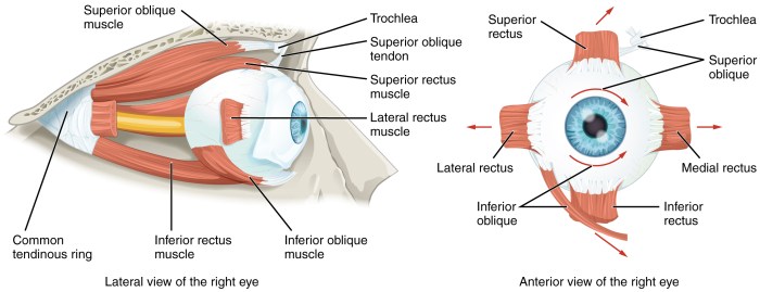
The extrinsic eye muscles are six muscles that control the movement of the eye. They are located outside the eyeball and are innervated by the oculomotor, trochlear, and abducens nerves.
Muscle Name, Origin, Insertion, Innervation, Action
The following table lists the six extrinsic eye muscles, their origin, insertion, innervation, and action:
| Muscle Name | Origin | Insertion | Innervation | Action |
|---|---|---|---|---|
| Superior rectus | Common tendinous ring | Superior surface of the eyeball | Oculomotor nerve | Elevates the eye |
| Inferior rectus | Common tendinous ring | Inferior surface of the eyeball | Oculomotor nerve | Depresses the eye |
| Medial rectus | Common tendinous ring | Medial surface of the eyeball | Oculomotor nerve | Adducts the eye |
| Lateral rectus | Common tendinous ring | Lateral surface of the eyeball | Abducens nerve | Abducts the eye |
| Superior oblique | Common tendinous ring | Trochlea | Trochlear nerve | Intorts the eye and depresses the ipsilateral eye and elevates the contralateral eye |
| Inferior oblique | Maxilla | Inferior surface of the eyeball | Oculomotor nerve | Extorts the eye and elevates the ipsilateral eye and depresses the contralateral eye |
Innervation of the Extrinsic Eye Muscles: Label The Extrinsic Eye Muscles.
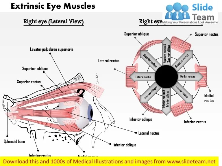
The extrinsic eye muscles are innervated by three cranial nerves: the oculomotor nerve (CN III), the trochlear nerve (CN IV), and the abducens nerve (CN VI).
The oculomotor nerve innervates all of the muscles that elevate, depress, and adduct the eye, as well as the muscle that constricts the pupil.
The trochlear nerve innervates the superior oblique muscle, which depresses and laterally rotates the eye.
The abducens nerve innervates the lateral rectus muscle, which abducts the eye.
| Muscle | Innervation |
|---|---|
| Superior rectus | Oculomotor nerve (CN III) |
| Inferior rectus | Oculomotor nerve (CN III) |
| Medial rectus | Oculomotor nerve (CN III) |
| Inferior oblique | Oculomotor nerve (CN III) |
| Superior oblique | Trochlear nerve (CN IV) |
| Lateral rectus | Abducens nerve (CN VI) |
Control of Eye Movements
The brainstem and cerebellum play crucial roles in controlling eye movements. The brainstem contains nuclei that innervate the extrinsic eye muscles, while the cerebellum coordinates and fine-tunes these movements.
Voluntary Eye Movements
Voluntary eye movements are initiated by the frontal eye fields in the cerebral cortex. These signals are sent to the brainstem, where they are processed by the superior colliculus and the nucleus of the oculomotor nerve (CN III). The superior colliculus is responsible for generating saccades, while the nucleus of CN III controls smooth pursuit movements.
Involuntary Eye Movements, Label the extrinsic eye muscles.
Involuntary eye movements are controlled by the brainstem and cerebellum. The vestibular nuclei in the brainstem detect head movements and generate compensatory eye movements to maintain visual fixation. The cerebellum coordinates these movements and fine-tunes their accuracy and speed.
Clinical Significance

The extrinsic eye muscles play a crucial role in vision and are responsible for controlling the movement and position of the eyes. Dysfunction or damage to these muscles can lead to various clinical conditions and visual impairments.
Common pathologies and dysfunctions associated with the extrinsic eye muscles include:
- Strabismus:Misalignment of the eyes, causing double vision (diplopia) and impaired depth perception.
- Ptosis:Drooping of the upper eyelid, obstructing vision.
- Nystagmus:Involuntary, rapid eye movements that can impair vision.
- Myasthenia gravis:A neuromuscular disorder that weakens the extrinsic eye muscles, leading to ptosis and diplopia.
- Third nerve palsy:Damage to the third cranial nerve, which innervates several extrinsic eye muscles, causing ptosis, diplopia, and impaired eye movement.
Clinical Tests for Extrinsic Eye Muscle Function
Several clinical tests are used to assess the function of the extrinsic eye muscles and diagnose related conditions:
- Cover test:Evaluates eye alignment by covering one eye and observing the movement of the uncovered eye.
- Slit-lamp examination:Uses a biomicroscope to examine the eye structures, including the extrinsic eye muscles.
- Forced duction test:Assesses muscle strength by manually moving the eye in different directions.
- Electronystagmography (ENG):Records eye movements to detect nystagmus and other abnormalities.
- Magnetic resonance imaging (MRI):Provides detailed images of the eye muscles and surrounding structures, helping diagnose conditions like third nerve palsy.
Answers to Common Questions
What are the six extrinsic eye muscles?
The six extrinsic eye muscles are the superior rectus, inferior rectus, medial rectus, lateral rectus, superior oblique, and inferior oblique.
Which cranial nerves innervate the extrinsic eye muscles?
The extrinsic eye muscles are innervated by three cranial nerves: the oculomotor nerve (CN III), the trochlear nerve (CN IV), and the abducens nerve (CN VI).
What is the function of the extrinsic eye muscles?
The extrinsic eye muscles control the movements of the eyes, allowing us to look up, down, left, right, and diagonally.
